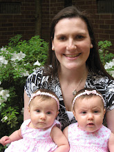
It was still early in the morning when one of the doctors came into the room with me to discuss Ruthie's MRI. She attempted to explain to me that her MRI results were not good. She had evidence of several "infarction's". I asked what an "infarction" was and basically she described it to me as a series of attacks on parts of her brain, similar to mini-strokes. It showed evidence of her being deprived of oxygen for extended periods of time. I asked if that had to do with the apnea episodes in the ER and she stated that it would have had to been longer than her episodes in the ER. I am sure she saw the look of pure confusion on my face and asked if I wanted to see the MRI's.

 Actually looking at them still made no sense to me. She "dumbed" it down for me to say that everywhere there are white marks on her MRI is brain damage. Those were words that made sense to me. I knew what brain damage was. What I didnt know what how my seemingly perfect 2 week old had brain damage.
 I was a psychology major in college, so we had to do a little bit of anatomy in regards to the brain. I knew that different parts of the brain controlled different things. I asked her what kinds of delays we should be expecting. Was the damage primarily in her motor skills section, her verbal comprehension, her reasoning, etc. The doctor told me it was too widespread to narrow it down to one area that she would be delayed in. I also knew about "re-mapping" which is where after damage occurs to the brain, some how it learns to "re-map" around the damaged area to learn how to do that function. I asked her since all she had "learned" how to do was eat, cry and poop, couldnt she just "re-map" how to learn the other things like crawling, rolling over, talking, math, etc. It seemed like a logical answer to me. She then told me that re-mapping can only occur when there is damage in one area, not in several areas as severe as hers. She just said that we would have to take one developmental milestone at a time and deal with it. She explained to me that I should not expect her to achieve any of the expected milestones, included simple things like holding her head up.
Before we were discharged from the hospital, they gave us CD's of the all of the MRI images. On another tab on the CD I discovered their "findings" and "impressions" that they attached to the results. Any one with a medical background will probably understand more of this than I do, but I get the gist of it.
MRV AND MR OF THE HEAD WITHOUT CONTRAST, 10/09/2007:
HISTORY: Seizures with history of apnea.
TECHNIQUE: Multiplanar, multisequence MR imaging of the brain is
performed without contrast. MRV is performed using TOF technique.
FINDINGS:
There is severely abnormal diffusion sequence with restricted
diffusion involving the majority of the subcortical white matter
including the corpus callus. Findings suggest a diffuse anoxic
injury. The cerebellum is spared. The cortex is spared. No
significant edema at this point. The dural venous sinuses are
patent.
IMPRESSION:
Severely abnormal MRI with diffuse infarct involving the majority of
the cerebral white matter.
END OF IMPRESSION
I was completely numb. I have twins, which is a handful in itself, and now I have a possibly handicap child to an extent that we dont even know. I was devastated and felt a pain to my core that only a mother with a sick child can understand. I let that feeling linger for about 5 minutes. Any one that knows me knows that I am a tad bit stubborn. I remember changing my prayers at that exact moment to asking God to help me with this challenge. Help me to be patient and do everything in my power to raise a handicapped child. It was going to be one milestone at a time, and I was going to do everything in my power and use EVERY resource available to me to make sure that we attacked each milestone head on.
Maybe that sounds a little cold, but to be completely honest, Mark was losing it (and he would not disagree with that statement), so some one had to buck up. I was Mommy. Mommy-mode kicked in, and I was going to take care of my child, no matter what her condition.
They decided to do an MRI on Maddie the next day to compare. After all, they were twins. They had been exposed to the same inter-utero conditions, same at home conditions, etc. Oh! And did I mention that since it looked like Ruthie had been deprived of oxygen, they were legally required to investigate for child abuse? So both girls were taken to get full body xrays to check for any broken bones, bruising, etc. Although I am glad for the precautions that are set in place to protect children, you cant help but be a little offended when they are inspecting your children for signs of child abuse after you have literally been camping out in a tiny room with your sick children, obviously distraught over the whole situation. Obviously no evidence was found that I had smothered them with a pillow, and they moved on to some other possible causes.
Maddie's MRI came back that same day.

 Same white spots. Same type of brain damage. Luckily it didnt seem as wide spread on Maddie's MRI as Ruthie's, but it was still brain damage. Here were the notes on Maddie's exam.
TECHNIQUE: Axial diffusion-weighted, T1-weighted, T2-weighted,
coronal T2 and FLAIR, and sagittal T1-weighted imaging were
performed.
FINDINGS:
Diffusion imaging reveals significant abnormal signal seen within the
subcortical white matter of the biparietal region and throughout the
splenium of the corpus callosum. Additionally, there is hyperintense
signal through the genu of the corpus callosum extending through the
minor forceps into the subcortical white matter of the frontal lobes.
Asymmetrically increased signal is also seen within the left centrum
semiovale. There is relative sparing of the posterior fossa and
brain stem structures.
Axial T1 imaging shows some vague scattered T1-hyperintense foci
within the bilateral centrum semiovale, left greater than right.
There is no mass effect. The ventricles are normal in configuration.
Coronal T2-weighted imaging shows no asymmetric morphology of the
temporal lobe regions.
IMPRESSION:
Significant abnormality involving the white matter as evident on the
diffusion sequence involving the periatrial white matter, corpus
callosum, deep and subcortical white matter of the bifrontal and
biparietal regions extending into the temporal lobes. There is
sparing of the brain stem and cerebellum. The findings likely
represent inborn error of metabolism, of uncertain pattern. The
pattern does appear very similar to the patient's sibling reported
separately.
Images and interpretation reviewed and verified by Dr. Wushensky.
END OF IMPRESSION
I was all of the sudden faced with the reality of having two handicapped children. That is not something that you can every truly be prepared for. Especially after a routine/boring pregnancy and un-eventful delivery. You just assume that when they tell you that they are perfect when they are born that it stays that way and that you have been through the worst of the uncertains. I had a million uncertains in front of me all of a sudden, and still no medical answers for me to hold on to. Looking back now, I think the lack of medical answers was God's way of forcing me to turn to him for answers to hold on to. He should always be the first one that I turn to, but lets face it, I am human and humans like facts and tangible things to hold on to. A diagnosis, a treatment plans. Any of those things I can hold on to and google. God knew what I needed more than what I thought I needed. He knew the end of the story already, but I was still in the storm.


1 comment:
Thank you for this post. Even in the midst of trial, your attitude is uplifting.
Post a Comment